Melanoma & Atypical Moles

Understanding the Deadliest Skin Cancer

Medical Dermatology
Overview

Melanoma is the most serious form of skin cancer because of its ability to spread to lymph nodes and distant organs. About 100,000 new invasive melanomas are diagnosed in the United States each year. The encouraging news is that when melanoma is caught early, while it is still confined to the skin, the five-year survival rate is around 99 percent. Late detection is what makes melanoma deadly, not the cancer itself.

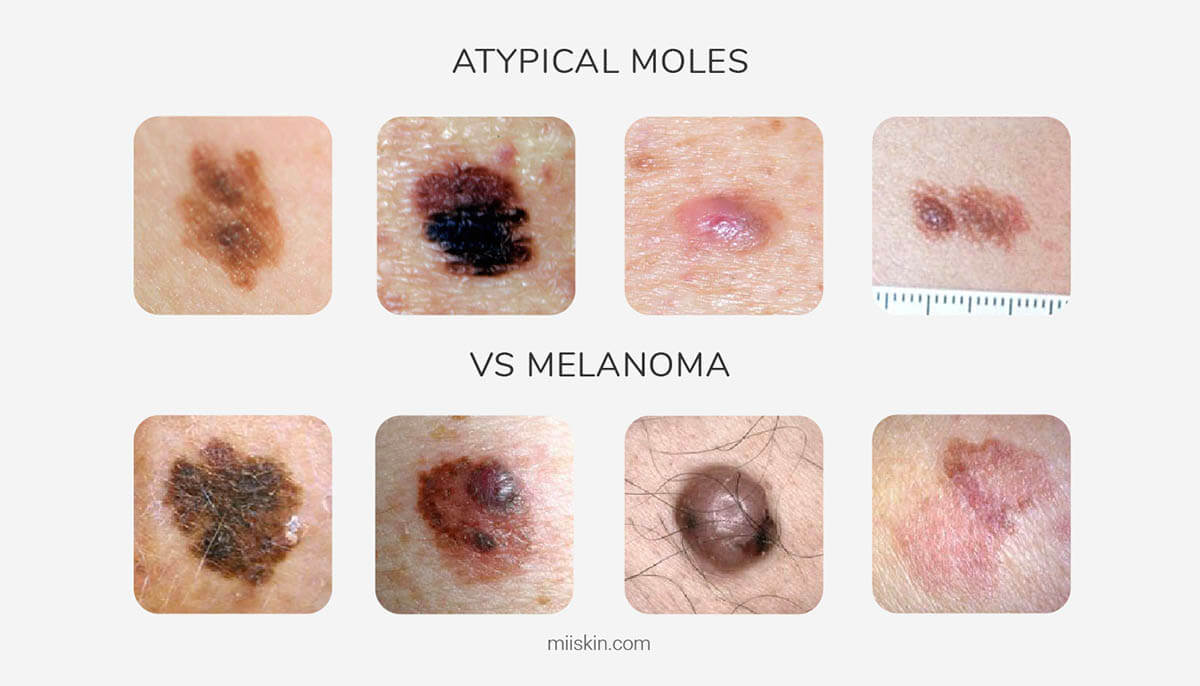
Atypical moles, also called dysplastic nevi, are unusual-looking moles that are not melanoma but do carry a higher risk of becoming melanoma over time and serve as a marker for elevated overall melanoma risk. People with multiple atypical moles, a personal or family history of melanoma, or both, benefit from regular dermatologic surveillance.

At Cleaver Dermatology and Aesthetics, our board-certified dermatologists provide full-body skin exams, dermoscopy, biopsy, and coordinated treatment for melanoma at all nine of our North Georgia and Metro Atlanta offices. We work closely with surgical oncology and medical oncology when advanced disease requires it.

Common Symptoms

The most useful framework for spotting melanoma is the ABCDE rule. A is for asymmetry: one half of the mole does not match the other. B is for border irregularity: the edges are notched, scalloped, or blurred. C is for color variation: shades of brown, black, red, white, or blue within a single lesion. D is for diameter greater than six millimeters, about the size of a pencil eraser, although melanomas can be smaller. E is for evolution: any change in size, shape, color, surface, or symptoms over time.

The ugly duckling sign is just as useful. If one mole on your body looks different from all your others, that is the one to show your dermatologist. Other warning signs include a new spot that appears in adulthood, itching or bleeding from a mole, a dark streak under a fingernail or toenail, and pigmented spots on the palms or soles. Acral and nodular melanomas can lack pigment and grow downward rapidly, so any rapidly growing pink or red bump on the skin also deserves attention.

Common Causes

Most melanomas are linked to ultraviolet radiation, particularly intense intermittent sun exposure and a history of blistering sunburns, especially in childhood. Tanning bed use before age 35 raises melanoma risk by about 75 percent. Some melanomas, however, arise on skin that gets little sun, which is why we examine the whole body, including the scalp, soles, and between the toes.

Personal risk factors include fair skin that burns easily, light eyes, blond or red hair, more than 50 moles, atypical moles, a personal history of any skin cancer, a family history of melanoma in a first-degree relative, immunosuppression, and certain inherited genetic mutations such as CDKN2A. Anyone with these factors should be on a regular skin exam schedule, and family members of patients with melanoma deserve their own evaluation.

Treatment Options

Treatment of melanoma is driven by stage. Early-stage melanoma confined to the skin is treated with wide local excision, which means removing the lesion plus a margin of healthy skin determined by tumor depth. For melanoma in situ, a five to ten millimeter margin is standard. For invasive melanoma, margins range from one to two centimeters depending on Breslow depth.

Sentinel lymph node biopsy is offered for melanomas with a Breslow depth of 0.8 millimeters or greater, or thinner lesions with high-risk features, to identify microscopic spread to the first draining lymph node. The result helps guide whether additional imaging or systemic therapy is needed.

For Stage III and Stage IV melanoma, treatment has been transformed by immunotherapy. Checkpoint inhibitors such as pembrolizumab and nivolumab, alone or combined with ipilimumab, produce durable responses in many patients. For tumors with a BRAF mutation, targeted therapy with combinations such as dabrafenib and trametinib offers another effective option. We coordinate this care with medical oncology partners.

Atypical moles that show concerning change are biopsied or excised. Severely atypical or moderate-to-severe dysplastic nevi with positive margins on biopsy are typically re-excised to remove the remaining cells.

What to Expect During Treatment

Your visit usually begins with a full-body skin exam from scalp to toes, using a dermatoscope to magnify lesions and look at vascular and pigment patterns invisible to the naked eye. We catalog concerning lesions, often photograph them for monitoring, and discuss whether biopsy is appropriate. Biopsy is a brief in-office procedure under local anesthesia.

Pathology results take five to seven business days. If melanoma is confirmed we walk you through Breslow depth, ulceration status, mitotic rate, and what those numbers mean for next steps. We schedule wide excision, coordinate sentinel node biopsy when indicated, and arrange staging imaging if the depth and features warrant it.

After treatment, follow-up includes total-body skin exams every three to six months for the first few years, then less often based on stage. Many patients benefit from total-body photography and serial dermoscopic monitoring, especially when atypical moles are part of the picture.

When to See a Dermatologist

Schedule an exam right away for any mole that meets the ABCDE criteria, the ugly duckling on your body, a new dark streak in a nail, a non-healing pigmented sore, or a mole that itches, bleeds, or changes. Any first-degree relative diagnosed with melanoma is reason for you to be evaluated even without a specific concern.

If you have many moles, atypical moles, fair skin with significant sun exposure, a history of tanning bed use, or are immunosuppressed, get on an annual or semi-annual skin exam schedule. Early detection truly changes outcomes.

Prevention and Self-Care

Daily broad-spectrum SPF 30 or higher, reapplied every two hours outdoors, paired with hats, sun shirts, and shade during peak hours, lowers your lifetime melanoma risk. Avoid tanning beds. Be especially protective of children, since blistering sunburns in childhood meaningfully raise adult melanoma risk.

Check your own skin once a month using a mirror, with help for the back and scalp. Photograph any mole you are watching, with a ruler in the frame for scale, so you can compare objectively over time. Pay attention to the new and the changing, not just the ugly. Then bring anything you are unsure about to your dermatologist promptly.

FAQs

How is melanoma different from other skin cancers?

Melanoma is the deadliest skin cancer because it spreads more readily than basal cell or squamous cell carcinoma. While BCC and SCC grow slowly and rarely spread, melanoma can metastasize to lymph nodes and distant organs even when relatively small. This metastatic potential makes early detection crucial. Melanoma can also develop in areas with minimal sun exposure, suggesting genetic factors play a more significant role than in other skin cancers.

What are atypical moles and why are they concerning?

Atypical moles, also called dysplastic nevi, are larger than normal moles with irregular borders and mixed colors. They carry increased risk for melanoma development. Patients with multiple atypical moles have significantly higher lifetime melanoma risk than the general population. At Cleaver Dermatology and Aesthetics, we monitor atypical moles closely and may recommend removal of lesions that are changing or have features suggesting malignancy.

What does the ABCDE rule mean?

The ABCDE rule helps you identify concerning moles. Asymmetry means one half doesn't match the other. Border irregularity means uneven edges. Color variation means multiple colors in one mole. Diameter greater than 6mm, roughly the size of a pencil eraser, is concerning. Evolution means any change over time. Any mole exhibiting these features warrants evaluation by a dermatologist.

Is early detection really important for melanoma?

Yes, early detection is absolutely critical for melanoma. Thin melanomas less than 1mm thick have 5-year survival rates exceeding 95 percent, while thick melanomas greater than 4mm have significantly poorer prognosis. Patients with melanomas detected before lymph node involvement have substantially better outcomes than those with advanced disease. This is why we emphasize monthly self-exams and annual professional screenings.

What happens after melanoma treatment?

After melanoma treatment, regular follow-up is essential to detect any recurrence early. You'll have frequent clinical skin exams, with frequency depending on your melanoma stage and risk factors. Some patients benefit from baseline imaging and periodic follow-up imaging. Lifelong sun protection and monthly self-examination are important. Most importantly, continue vigilant skin monitoring for new or changing lesions.

Menu